World Journal of Emergency Medicine ›› 2022, Vol. 13 ›› Issue (3): 175-181.doi: 10.5847/wjem.j.1920-8642.2022.057
• Original Articles • Previous Articles Next Articles
Xuan Fu1, Xue Lin2, Samuel Seery3, Li-na Zhao1, Hua-dong Zhu1, Jun Xu1( ), Xue-zhong Yu1()
Received:2022-02-10
Accepted:2022-04-20
Online:2022-05-13
Published:2022-05-01
Contact:
Jun Xu,Xue-zhong Yu
E-mail:xujunfree@126.com;yxzpumch@126.com
Xuan Fu, Xue Lin, Samuel Seery, Li-na Zhao, Hua-dong Zhu, Jun Xu, Xue-zhong Yu. Speckle-tracking echocardiography for detecting myocardial dysfunction in sepsis and septic shock patients: A single emergency department study[J]. World Journal of Emergency Medicine, 2022, 13(3): 175-181.
Add to citation manager EndNote|Ris|BibTeX
URL: http://wjem.com.cn/EN/10.5847/wjem.j.1920-8642.2022.057
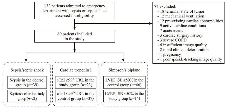
Table 1.
Two-dimensional echocardiographic parameters and laboratory tests of the investigated groups
| Variables | Sepsis 3.0 | cTnI (99th URL) 0.056 μg/L | LVEF_SB | ||||||
|---|---|---|---|---|---|---|---|---|---|
| Septic shock (n=21) | Sepsis (n=39) | P | ≥99th URL (n=23) | <99th URL (n=37) | P | <50% (n=14) | ≥50% (n=46) | P | |
| Conventional echocardiography | |||||||||
| LVEF_Teich, % | 61.45±11.55 | 62.53±7.43 | 0.70 | 58.97±9.45 | 64.09±8.28 | 0.03* | 54.49±9.50 | 64.45±7.55 | 0.01* |
| LVEF_SB, % | 53.42±6.96 | 55.04±6.19 | 0.36 | 51.71±7.59 | 56.17±5.06 | 0.01* | 45.79±3.82 | 57.10±4.49 | 0.01* |
| CI, % | 29.88±16.59 | 28.81±12.76 | 0.77 | 28.39±14.46 | 29.68±14.09 | 0.73 | 0.27±0.16 | 0.30±0.13 | 0.49 |
| LVOT_VTI, mm | 16.52±3.94 | 17.22±3.59 | 0.50 | 15.74±3.80 | 17.74±3.46 | 0.04* | 14.63±3.72 | 17.69±3.41 | 0.01* |
| LVOT_SV, mL | 39.34±11.69 | 44.51±10.26 | 0.08 | 38.87±11.05 | 45.08±10.34 | 0.03* | 40.38±13.50 | 43.40±10.13 | 0.37 |
| LVOT_CO, L/min | 3.83±1.42 | 3.85±1.24 | 0.93 | 3.83±1.49 | 3.78±1.15 | 0.92 | 3.74±1.40 | 3.82±1.26 | 0.83 |
| E/A | 1.01±0.26 | 1.07±0.35 | 0.33 | 1 (0.90, 1.18) | 1 (0.94, 1.17) | 0.58 | 1 (1.00, 1.55) | 1 (0.91, 1.14) | 0.26 |
| e', cm/s | 12.48±3.84 | 12.12±3.04 | 0.69 | 12.23±3.51 | 12.23±3.20 | 0.95 | 12.55±4.20 | 12.13±3.01 | 0.68 |
| E/e' ratio (lateral) | 6.33±2.19 | 6.02±2.07 | 0.59 | 6.13±2.42 | 6.07±1.88 | 0.77 | 5.89±2.32 | 6.16±2.03 | 0.68 |
| Speckle-tracking echocardiography | |||||||||
| GLS, % | -15.48±3.43 | -16.59±3.82 | 0.27 | -14.74±4.21 | -17.11±3.06 | 0.02* | -12.29±2.61 | -17.39±3.12 | 0.01* |
| GCS, % | -17.19±4.77 | -16.38±4.17 | 0.50 | -15.17±4.31 | -17.59±4.19 | 0.04* | -14.14±4.00 | -17.43±4.22 | 0.01* |
| GRS, % | 22.81±5.68 | 19.87±5.56 | 0.06 | 19.35±5.89 | 21.86±5.49 | 0.09 | 18.79±5.40 | 21.54±5.73 | 0.12 |
| LVEF_STI, % | 49.00±10.81 | 50.78±9.59 | 0.52 | 46.61±11.37 | 52.36±8.43 | 0.03* | 38.31±8.25 | 53.76±7.34 | 0.01* |
| Laboratory test | |||||||||
| Platelet, ×109/L | 186.05±96.42 | 178.85±100.07 | 0.79 | 149.61±88.79 | 201.11±99.47 | 0.05 | 132.00±86.25 | 196.39±97.30 | 0.03* |
| CRP, mg/L | 127.48±89.94 | 152.93±81.46 | 0.64 | 150.67±82.50 | 160.19±80.60 | 0.66 | 171.75±87.04 | 151.91±79.18 | 0.43 |
| Procalcitonin, ng/L | 4.65 (0.58, 16.50) | 6.00 (1.00, 16.00) | 0.17 | 10.00 (2.90, 74.00) | 5.30 (1.40, 18.00) | 0.04* | 10.00 (5.25, 91.00) | 5.20 (1.30, 17.00) | 0.14 |
| cTnI, ng/mL | 0.03 (0, 0.68) | 0.02 (0, 0.02) | 0.23 | 0 (0, 3.00) | 0.02 (0, 0.02) | 0.01* | 0.02 (0, 3.25) | 0.02 (0, 0.02) | 0.16 |
| NT-proBNP, ng/mL | 2989 (1002, 9663) | 1031 (165, 5423) | 0.02* | 6436 (2763, 17254) | 881 (200, 1596) | 0.01* | 12026 (4929, 18810) | 1028 (246, 2832) | 0.01* |
| CK, U/L | 108 (68, 391) | 72 (34, 271) | 0.31 | 180 (74, 414) | 63 (32, 136) | 0.30 | 233 (61, 945) | 75 (35, 248) | 0.11 |
| CK-MB, U/L | 1.8 (0.8, 5.7) | 1.0 (0, 2.0) | 0.05 | 2.0 (1.0, 9.0) | 1.0 (0, 2.0) | 0.10 | 2.0 (1.0, 18.0) | 1.0 (0, 2.1) | 0.02* |
| Cr, μmol/L | 156 (106, 224) | 87 (59, 139) | 0.01* | 136 (94, 209) | 95 (58, 150) | 0.05 | 159 (82, 232) | 103 (63, 146) | 0.09 |
| Lac, mmol/L | 3.3 (2.2,5.4) | 2.0 (1.0, 2.0) | 0.01* | 2.0 (2.0, 4.0) | 2.0 (1.0, 3.0) | 0.29 | 2.5 (2.0, 5.3) | 2.0 (1.0, 3.0) | 0.09 |
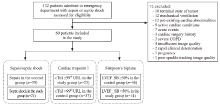
Figure 1.
Flowchart for participant assignment. cTnI: cardiac troponin I; LVEF: left ventricular ejection fraction; SB: Simpson's biplane; COPD: chronic obstructive pulmonary disease; 99th URL: 99 percentile upper reference limit.
| 1 |
Hollenberg SM, Singer M. Pathophysiology of sepsis-induced cardiomyopathy. Nat Rev Cardiol. 2021; 18(6):424-34.
doi: 10.1038/s41569-020-00492-2 pmid: 33473203 |
| 2 |
Beesley SJ, Weber G, Sarge T, Nikravan S, Grissom CK, Lanspa MJ, et al. Septic cardiomyopathy. Crit Care Med. 2018; 46(4):625-34.
doi: 10.1097/CCM.0000000000002851 pmid: 29227368 |
| 3 |
Lin H, Wang WT, Lee M, Meng QH, Ren HS. Current status of septic cardiomyopathy: basic science and clinical progress. Front Pharmacol. 2020; 11:210.
doi: 10.3389/fphar.2020.00210 |
| 4 |
Voigt JU, Pedrizzetti G, Lysyansky P, Marwick TH, Houle H, Baumann R, et al. Definitions for a common standard for 2D speckle tracking echocardiography: consensus document of the EACVI/ASE/Industry task force to standardize deformation imaging. Eur Heart J Cardiovasc Imaging. 2015; 16(1):1-11.
doi: 10.1093/ehjci/jeu184 |
| 5 |
Houard L, Militaru S, Tanaka K, Pasquet A, Vancraeynest D, Vanoverschelde JL, et al. Test-retest reliability of left and right ventricular systolic function by new and conventional echocardiographic and cardiac magnetic resonance parameters. Eur Heart J Cardiovasc Imaging. 2020; 22(10):1157-67.
doi: 10.1093/ehjci/jeaa206 |
| 6 | Onishi T, Saha SK, Delgado-Montero A, Ludwig DR, Onishi T, Schelbert EB, et al. Global longitudinal strain and global circumferential strain by speckle-tracking echocardiography and feature-tracking cardiac magnetic resonance imaging: comparison with left ventricular ejection fraction. J Am Soc Echocardiogr. 2015; 28(5):587-96. |
| 7 |
Cai AP, Zhu YC, Clark SA, Feng YQ. The use of machine learning for the care of hypertension and heart failure. JACC: Asia. 2021; 1(2):162-72.
doi: 10.1016/j.jacasi.2021.07.005 |
| 8 |
Orde SR, Pulido JN, Masaki M, Gillespie S, Spoon JN, Kane GC, et al. Outcome prediction in sepsis: speckle tracking echocardiography based assessment of myocardial function. Crit Care. 2014; 18(4):R149.
doi: 10.1186/cc13987 |
| 9 |
Ng PY, Sin WC, Ng AK, Chan WM. Speckle tracking echocardiography in patients with septic shock: a case control study (SPECKSS). Crit Care. 2016; 20(1):145.
doi: 10.1186/s13054-016-1327-0 |
| 10 | Hai PD, Binh NT, Hien NVQ, Hoang NH, Hoan VN, Son PN, et al. Prognostic role of left ventricular systolic function measured by speckle tracking echocardiography in septic shock. Biomed Res Int. 2020; 2020:7927353. |
| 11 |
Sanfilippo F, Corredor C, Fletcher N, Tritapepe L, Lorini FL, Arcadipane A, et al. Left ventricular systolic function evaluated by strain echocardiography and relationship with mortality in patients with severe sepsis or septic shock: a systematic review and meta-analysis. Crit Care. 2018; 22(1):183.
doi: 10.1186/s13054-018-2113-y |
| 12 |
Vallabhajosyula S, Rayes HA, Sakhuja A, Murad MH, Geske JB, Jentzer JC. Global longitudinal strain using speckle-tracking echocardiography as a mortality predictor in sepsis: a systematic review. J Intensive Care Med. 2019; 34(2):87-93.
doi: 10.1177/0885066618761750 pmid: 29552957 |
| 13 |
Singer M, Deutschman CS, Seymour CW, Shankar-Hari M, Annane D, Bauer M, et al. The third international consensus definitions for sepsis and septic shock (Sepsis 3.0). JAMA. 2016; 315(8):801-10.
doi: 10.1001/jama.2016.0287 |
| 14 |
Evans L, Rhodes A, Alhazzani W, Antonelli M, Coopersmith CM, French C, et al. Surviving Sepsis Campaign: international guidelines for management of sepsis and septic shock 2021. Crit Care Med. 2021; 49(11):e1063-143.
doi: 10.1097/CCM.0000000000005337 |
| 15 |
Lang RM, Badano LP, Mor-Avi V, Afilalo J, Armstrong A, Ernande L, et al. Recommendations for cardiac chamber quantification by echocardiography in adults: an update from the American Society of Echocardiography and the European Association of Cardiovascular Imaging. J Am Soc Echocardiogr. 2015; 28(1):1-39.e14.
doi: 10.1016/j.echo.2014.10.003 |
| 16 |
Shahul S, Gulati G, Hacker MR, Mahmood F, Canelli R, Nizamuddin J, et al. Detection of myocardial dysfunction in septic shock: a speckle-tracking echocardiography study. Anesth Analg. 2015; 121(6):1547-54.
doi: 10.1213/ANE.0000000000000943 pmid: 26397444 |
| 17 |
Lanspa MJ, Shahul S, Hersh A, Wilson EL, Olsen TD, Hirshberg EL, et al. Associations among left ventricular systolic function, tachycardia, and cardiac preload in septic patients. Ann Intensive Care. 2017; 7(1):17.
doi: 10.1186/s13613-017-0240-2 |
| 18 |
Innocenti F, Palmieri V, Stefanone VT, D'Argenzio F, Cigana M, Montuori M, et al. Prognostic stratification in septic patients with overt and cryptic shock by speckle tracking echocardiography. Intern Emerg Med. 2021; 16(3):757-64.
doi: 10.1007/s11739-020-02545-3 |
| 19 |
Cameli M, Mandoli GE, Sciaccaluga C, Mondillo S. More than 10 years of speckle tracking echocardiography: still a novel technique or a definite tool for clinical practice? Echocardiography. 2019; 36(5):958-70.
doi: 10.1111/echo.14339 |
| 20 |
Potter E, Marwick TH. Assessment of left ventricular function by echocardiography: the case for routinely adding global longitudinal strain to ejection fraction. JACC Cardiovasc Imaging. 2018; 11(2 Pt 1):260-74.
doi: 10.1016/j.jcmg.2017.11.017 |
| 21 | Li T, Liu JJ, Du WH, Wang X, Chen ZQ, Zhang LC. 2D speckle tracking imaging to assess sepsis induced early systolic myocardial dysfunction and its underlying mechanisms. Eur Rev Med Pharmacol Sci. 2014; 18(20):3105-14. |
| 22 |
Hestenes SM, Halvorsen PS, Skulstad H, Remme EW, Espinoza A, Hyler S, et al. Advantages of strain echocardiography in assessment of myocardial function in severe sepsis: an experimental study. Crit Care Med. 2014; 42(6):e432-40.
doi: 10.1097/CCM.0000000000000310 |
| 23 |
de Abreu CB, Muzzi RAL, Schulien T, Coelho MR, Alves LA, et al. Systolic dysfunction by two-dimensional speckle tracking echocardiography in dogs with parvoviral enteritis. J Vet Cardiol. 2021; 34:93-104.
doi: 10.1016/j.jvc.2021.01.006 |
| 24 |
Farsalinos KE, Daraban AM, Ünlü S, Thomas JD, Badano LP, Voigt JU. Head-to-head comparison of global longitudinal strain measurements among nine different vendors: the EACVI/ASE inter-vendor comparison study. J Am Soc Echocardiogr. 2015; 28(10):1171-81, e2.
doi: 10.1016/j.echo.2015.06.011 |
| 25 |
Innocenti F, Palmieri V, Guzzo A, Stefanone VT, Donnini C, Pini R. SOFA score and left ventricular systolic function as predictors of short-term outcome in patients with sepsis. Intern Emerg Med. 2018; 13(1):51-8.
doi: 10.1007/s11739-016-1579-3 |
| 26 |
Innocenti F, Palmieri V, Stefanone VT, D'Argenzio F, Cigana M, Montuori M, et al. Comparison of Troponin I levels versus myocardial dysfunction on prognosis in sepsis. Intern Emerg Med. 2022; 17(1):223-31.
doi: 10.1007/s11739-021-02701-3 |
| 27 |
Dalla K, Hallman C, Bech-Hanssen O, Haney M, Ricksten SE. Strain echocardiography identifies impaired longitudinal systolic function in patients with septic shock and preserved ejection fraction. Cardiovasc Ultrasound. 2015; 13:30.
doi: 10.1186/s12947-015-0025-4 |
| 28 |
Zaky A, Gill EA, Lin CP, Paul CP, Bendjelid K, Treggiari MM. Characteristics of Sepsis-induced cardiac dysfunction using speckle-tracking echocardiography: a feasibility study. Anaesth Intensive Care. 2016; 44(1):65-76.
doi: 10.1177/0310057X1604400111 |
| 29 |
Masson S, Caironi P, Fanizza C, Carrer S, Caricato A, Fassini P, et al. Sequential N-terminal pro-B-type natriuretic peptide and high-sensitivity cardiac troponin measurements during albumin replacement in patients with severe sepsis or septic shock. Crit Care Med. 2016; 44(4):707-16.
doi: 10.1097/CCM.0000000000001473 pmid: 26571184 |
| [1] | Mei-jia Shen, Li-chao Sun, Xiao-yu Liu, Meng-chen Xiong, Shan Li, A-ling Tang, Guo-qiang Zhang. Trichostatin A improves the inflammatory response and liver injury in septic mice through the FoxO3a/autophagy signaling pathway [J]. World Journal of Emergency Medicine, 2022, 13(3): 182-188. |
| [2] | Hai Hu, Jing-yuan Jiang, Ni Yao. Comparison of different versions of the quick sequential organ failure assessment for predicting in-hospital mortality of sepsis patients: A retrospective observational study [J]. World Journal of Emergency Medicine, 2022, 13(2): 114-119. |
| [3] | Ren-qi Yao, Chao Ren, Di Ren, Jin-xiu Li, Ying Li, Xue-yan Liu, Lei Huang, Yong Liu, Mian Peng, Yong-wen Feng, Yong-ming Yao. Development of septic shock and prognostic assessment in critically ill patients with coronavirus disease outside Wuhan, China [J]. World Journal of Emergency Medicine, 2021, 12(4): 293-298. |
| [4] | Xin Lu, Wei Han, Yan-xia Gao, Shi-gong Guo, Shi-yuan Yu, Xue-zhong Yu, Hua-dong Zhu, Yi Li. Efficacy and safety of corticosteroids in immunocompetent patients with septic shock [J]. World Journal of Emergency Medicine, 2021, 12(2): 124-130. |
| [5] | Li-wei Duan, Jin-long Qu, Jian Wan, Yong-hua Xu, Yi Shan, Li-xue Wu, Jin-hao Zheng, Wei-wei Jiang, Qi-tong Chen, Yan Zhu, Jian Zhou, Wen-bo Yu, Lei Pei, Xi Song, Wen-fang Li, Zhao-fen Lin. Effects of viral infection and microbial diversity on patients with sepsis: A retrospective study based on metagenomic next-generation sequencing [J]. World Journal of Emergency Medicine, 2021, 12(1): 29-35. |
| [6] | Hai-jiang Zhou, Tian-fei Lan, Shu-bin Guo. Outcome prediction value of National Early Warning Score in septic patients with community-acquired pneumonia in emergency department: A single-center retrospective cohort study [J]. World Journal of Emergency Medicine, 2020, 11(4): 206-215. |
| [7] | Yu-ming Wang, Yan-jun Zheng, Ying Chen, Yun-chuan Huang, Wei-wei Chen, Ran Ji, Li-li Xu, Zhi-tao Yang, Hui-qiu Sheng, Hong-ping Qu, En-qiang Mao, Er-zhen Chen. Effects of fluid balance on prognosis of acute respiratory distress syndrome patients secondary to sepsis [J]. World Journal of Emergency Medicine, 2020, 11(4): 216-222. |
| [8] | Miao Yuan, Ding-yi Yan, Fang-shi Xu, Yi-di Zhao, Yang Zhou, Long-fei Pan. Effects of sepsis on hippocampal volume and memory function [J]. World Journal of Emergency Medicine, 2020, 11(4): 223-230. |
| [9] | Wen-peng Yin, Jia-bao Li, Xiao-fang Zheng, Le An, Huan Shao, Chun-sheng Li. Effect of neutrophil CD64 for diagnosing sepsis in emergency department [J]. World Journal of Emergency Medicine, 2020, 11(2): 79-86. |
| [10] | Shao-hua Liu, Huo-yan Liang, Hong-yi Li, Xian-fei Ding, Tong-wen Sun, Jing Wang. Effect of low high-density lipoprotein levels on mortality of septic patients: A systematic review and meta-analysis of cohort studies [J]. World Journal of Emergency Medicine, 2020, 11(2): 109-116. |
| [11] | Yi-wen Fan, Shao-wei Jiang, Jia-meng Chen, Hui-qi Wang, Dan Liu, Shu-ming Pan, Cheng-jin Gao. A pulmonary source of infection in patients with sepsis-associated acute kidney injury leads to a worse outcome and poor recovery of kidney function [J]. World Journal of Emergency Medicine, 2020, 11(1): 18-26. |
| [12] | Kimberly A. Chambers, Adam Y. Park, Rosa C. Banuelos, Bryan F. Darger, Bindu H. Akkanti, Annamaria Macaluso, Manoj Thangam, Pratik B. Doshi. Outcomes of severe sepsis and septic shock patients after stratification by initial lactate value [J]. World Journal of Emergency Medicine, 2018, 9(2): 113-117. |
| [13] | Yan Ma, Xiang-you Yu, Yi Wang. Dose-related effects of dexmedetomidine on immunomodulation and mortality to septic shock in rats [J]. World Journal of Emergency Medicine, 2018, 9(1): 56-63. |
| [14] | Muhammad Akbar Baig, Hira Shahzad, Erfan Hussain, Asad Mian. Validating a point of care lactate meter in adult patients with sepsis presenting to the emergency department of a tertiary care hospital of a low- to middle-income country [J]. World Journal of Emergency Medicine, 2017, 8(3): 184-189. |
| [15] | Liang-shan Peng, Juan Li, Gao-sheng Zhou, Lie-hua Deng, Hua-guo Yao. Relationships between genetic polymorphisms of triggering receptor expressed on myeloid cells-1 and septic shock in a Chinese Han population [J]. World Journal of Emergency Medicine, 2015, 6(2): 123-130. |
| Viewed | ||||||
|
Full text |
|
|||||
|
Abstract |
|
|||||
