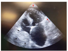
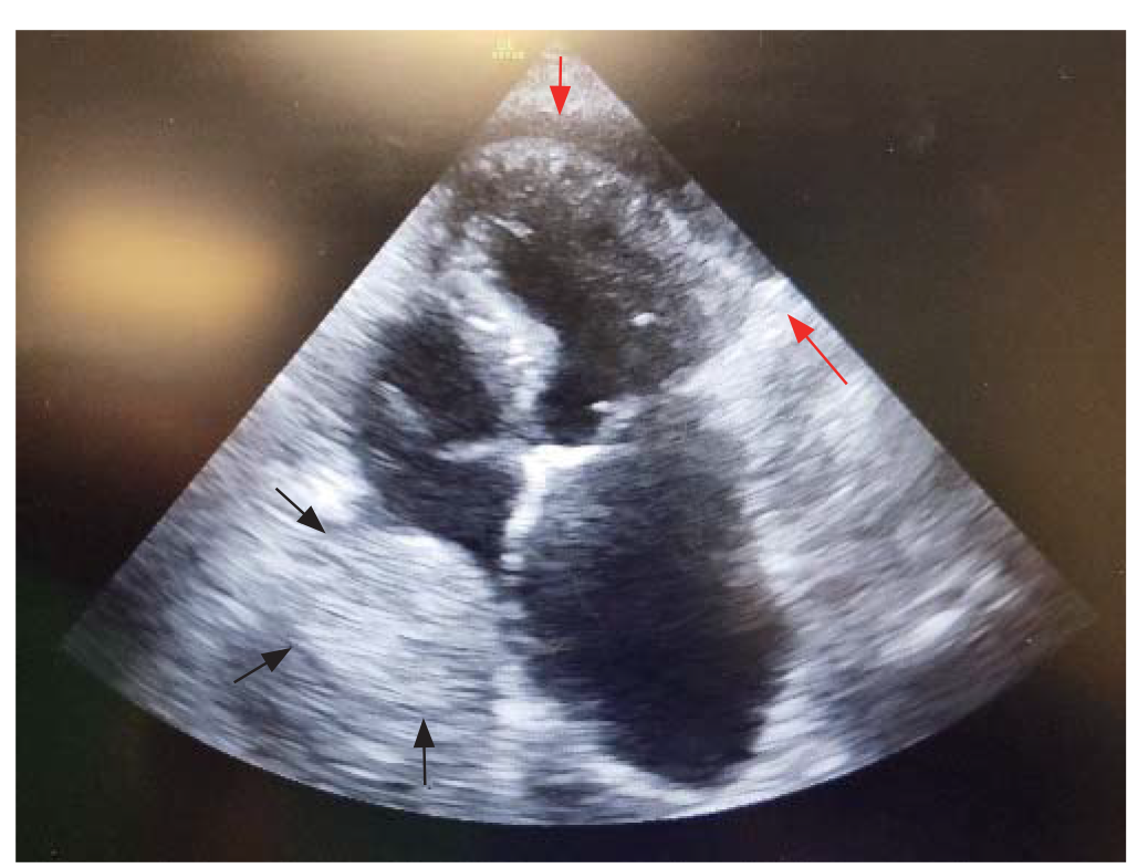
FoCUS apical four-chamber view showing small pericardial effusion (red arrows) and a round hematoma compressing the right atrium (black arrows). FoCUS: focused cardiac ultrasound.
Figure 1.
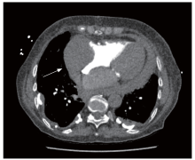
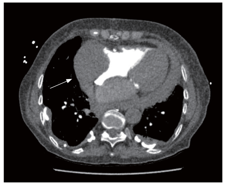
Figure 2.
The chest CT showed significant irregular wall thickening of all the cardiac chambers, especially the right atrium, with bulging into the lumen (white arrow). CT: computed tomography.
Hamdan M, Khoury F, Kossaify A. Loculated pericardial hematoma compressing the right atrium post mechanical aortic valve replacement and the role of point-of-care echocardiography: a case report. J Med Case Rep. 2023; 17(1):264.
2
Alerhand S, Adrian RJ, Long B, Avila J. Pericardial tamponade: a comprehensive emergency medicine and echocardiography review. Am J Emerg Med. 2022; 58:159-174.
doi: 10.1016/j.ajem.2022.05.001
pmid: 35696801
3
Arvig MD, Weile JB, Lindberg M, Wamberg J, Posth S. Focused cardiac ultrasound in emergency medicine. Ugeskr Laeger. 2023; 185(25):V02230130. [Article in Danish].

 ), Esther Montoro2, Leandro Noblia2