World Journal of Emergency Medicine ›› 2012, Vol. 3 ›› Issue (3): 213-220.doi: 10.5847/wjem.j.issn.1920-8642.2012.03.010
• Original Articles • Previous Articles Next Articles
Ying-zhen Wang1,2, Shi-wen Wang1( ), You-cheng Zhang2, Zhi-jiang Sun3
Received:2012-04-06
Accepted:2012-07-20
Online:2012-09-15
Published:2012-09-15
Contact:
Shi-wen Wang
E-mail:wshw120@163.com
Ying-zhen Wang, Shi-wen Wang, You-cheng Zhang, Zhi-jiang Sun. Protective effect of exogenous IGF-I on the intestinal mucosal barrier in rats with severe acute pancreatitis[J]. World Journal of Emergency Medicine, 2012, 3(3): 213-220.
Add to citation manager EndNote|Ris|BibTeX
URL: http://wjem.com.cn//EN/10.5847/wjem.j.issn.1920-8642.2012.03.010

Figure 1.
Changes of the serum amylase level in three groups.Compared with the SO group, *P<0.05, #P<0.01; compared with the SAP group, ΔP<0.05, ☆P<0.01.
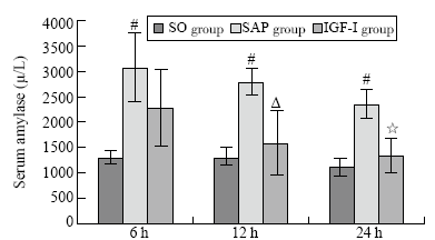

Figure 2.
Changes of the serum endotoxin level in the three groups.Compared with the SO group, *P<0.05, #P<0.01; compared with the SAP group, ΔP<0.05, ☆P<0.01.
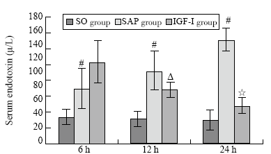

Figure 3.
Changes of the serum diamine oxidase level in the three groups. Compared with the SO group, *P<0.05 and #P<0.01; compared with the SAP group, ΔP<0.05, ☆P<0.01.
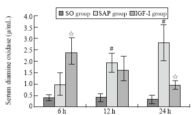
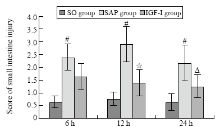
Figure 4.
Changes of score after small intestine injury in the three groups. Compared with the SO group, *P<0.05, #P<0.01; compared with the SAP group, ΔP<0.05, ☆P<0.01.
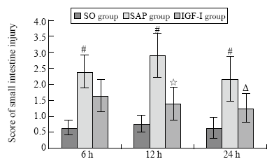

Figure 5.
Changes of the apoptotic index in the three groups. Compared with the SO group, *P<0.05, #P<0.01; compared with the SAP group, ΔP<0.05, ☆P<0.01.
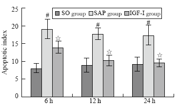
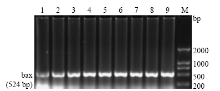
Figure 6.
The expression of bax mRNA.

Table 1
Changes of the expression of bax, bcl-2 mRNA and bax / bcl-2 in the three groups (mean ±SD)
| Groups | bax | bcl-2 | bax / bcl-2 | ||||||||
|---|---|---|---|---|---|---|---|---|---|---|---|
| 6 h | 12 h | 24 h | 6 h | 12 h | 24 h | 6 h | 12 h | 24 h | |||
| SO | 0.85±0.04 | 0.86±0.03 | 1.05±0.03 | 0.54±0.04 | 0.56±0.05 | 0.54±0.03 | 1.59±0.14 | 1.55±0.15 | 1.55±0.11 | ||
| SAP | 1.19±0.04# | 1.16±0.02# | 1.14±0.03* | 0.57±0.02 | 0.57±0.01 | 0.58±0.01 | 2.24±0.11# | 2.10±0.10# | 2.06±0.16# | ||
| IGF-I | 1.10±0.02 | 0.97±0.04Δ | 0.91±0.03Δ | 0.65±0.02☆ | 0.69±0.04▲ | 0.72±0.02☆ | 1.69±0.08☆ | 1.41±0.11☆ | 1.21±0.07☆ | ||
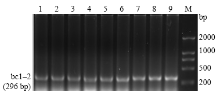
Figure 7.
The expression of bcl-2 mRNA.

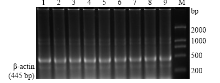
Figure 8.
The expression of β-actin mRNA.

| 1 | Xu GF, Lu Z, Gao J, Li ZS, Gong YF. Effect of ecoimmun-onutrition supports on maintenance of integrity of intestinal mucosal barrier in severe acute pancreatitis in dogs. Chin Med J (Engl) 2006; 119:656-661. |
| 2 |
Leveau P, Wang X, Sun Z, Börjesson A, Andersson E, Andersson R. Severity of pancreatitis-associated gut barrier dysfunction is reduced following treatment with the PAF inhibitor lexipafant. Biochem Pharmacol 2005; 69:1325-1331.
doi: 10.1016/j.bcp.2005.01.023 pmid: 15826603 |
| 3 | Sun B, Dong CG, Wang G, Jiang HC, Meng QH, Li J. et al. Analysis of fatal risk factors for severe acute pancreatitis: a report of 141 cases. Zhonghua Wai Ke Zazhi 2007; 45:1619-1622. |
| 4 |
Barreto S, Rodrigues J. Acute pancreatitis in Goa-A hospital-based study. J Indian Med Assoc 2008; 106:575-578.
pmid: 19552084 |
| 5 |
Barreto S, Rodrigues J. Comparison of Apache II and Imrie scoring systems in predicting the severity of acute pancreatitis. World J Emerg Surg 2007; 32-33.
pmid: 17076896 |
| 6 |
Sha H, Ma Q, Jha RK. Trypsin is the culprit of multiple organ injury with severe acute pancreatitis. Med Hypotheses 2009; 72:180-182.
doi: 10.1016/j.mehy.2008.09.007 pmid: 18938042 |
| 7 |
Czako L, Hegyi P, Takacs T. Effects of octreotide on acute necrotizing pancreat it is in rabbits. World J Gastroenterol 2004; 10:2082-2086.
doi: 10.3748/wjg.v10.i14.2082 pmid: 15237439 |
| 8 |
Telek G, Regöly-Mérei J, Kovács GC, Simon L, Nagy Z, Hamar J. et al. The first histological demonstration of ancreatic oxidative stress in human acute pancreatitis. Hepatogastroenterology 2001; 48:1252-1258.
pmid: 11677940 |
| 9 |
Baregamian N, Song J, Jeschke MG, Evers BM, Chung DH. IGF-I protects intestinal epithelial cells from oxidative stress-induced apoptosis. J Surg Res 2006; 136:31-37.
doi: 10.1016/j.jss.2006.04.028 pmid: 16999977 |
| 10 |
Nobumichi H, Masaharu N, Manabtu N, Hiramatsu Y, Hioki K, Yamamoto M. Structural and functional alterations in the gut of parenterally or enterally fed rats. J Surg Res 1989; 47:129-132.
doi: 10.1016/0022-4804(89)90076-0 pmid: 2547111 |
| 11 |
Chiu CJ, Mcardle AH, Brown R, Scott HJ, Gurd FN. Intestinal mucosal lesions in low flow states. I. A morphological, hemodynamic, and metabolic reappraisal. Arch Surg 1970; 101:478-483.
pmid: 5457245 |
| 12 |
Van Felius ID, Akkermans LM, Bosscha K, Verheem A, Harmsen W, Visser MR. et al. Interdigestive small bowel motility and duodenal bacterial overgrowth in experimental acute pancreatitis. Neurogastroenterol Motil 2003; 15:267-276.
doi: 10.1046/j.1365-2982.2003.00410.x pmid: 12787336 |
| 13 |
Gavirele Y, Sherman Y, Ben-Sasson SA. Identification of programmed cell death in situ via specific labeling of nuclear DNA fragmentation. J Cell Biol 1992; 119:493-501.
pmid: 1400587 |
| 14 | Wang LC, Ma QY, Sha HC. The effects of resveratrol on calcium regulation in rats with severe acute pancreatitis. Chin J Gen Surg 2006; 20:809-810. |
| 15 | Butturini G, Salvia R, Bettini R, Falconi M, Pederzoli P, Bassi C. Infection prevention in necrotizing pancreatitis: an old challenge with new perspectives. J Hosp Infect 2001; 49:4281. |
| 16 |
Matsuda N, Nishihira J, Takahashi Y, Kemmotsu O, Hattori Y. Role of macrophage migration inhibitory factor in acute lung injury in mice with acute pancreatitis complicated by endotoxemia. Am J Respir Cell Mol Biol 2006; 35:198-205.
pmid: 16574946 |
| 17 |
Qiao SF, Lu TJ, Sun JB, Li F. Alterations of intestinal immune function and regulatory effects of L-arginine in experimental severe acute pancreatitis rats. World J Gastroenterol 2005; 11:6216-6218.
doi: 10.3748/wjg.v11.i39.6216 pmid: 16273654 |
| 18 |
Marotta F, Barreto R, Wu CC, Naito Y, Gelosa F, Lorenzetti A. et al. Experimental acute alcohol pancreatitis-related liver damage and endotoxemia: synbiotics but not metronidazole have a protective effect. Chin J Dig Dis 2005; 6:193-197.
pmid: 16246229 |
| 19 |
Yasuda T, Takeyama Y, Ueda T, Shinzeki M, Sawa H, Nakajima T. et al. Break down of intestinal mucosa via accelerated apoptosis increases intestinal permeability in experimental severe acute pancreatitis. J Surg Res 2006; 135:18-26.
doi: 10.1016/j.jss.2006.02.050 pmid: 16603187 |
| 20 | Coopersmith CM, Chang KC, Swanson PE, Tinsley KW, Stromberg PE, Buchman TG. et al. Overexpression of Bc1-2 in the intestinal epithelium improves survival in septic mice. Care Med 2002; 30:l95-201. |
| 21 |
Cui J, Engelman RM, Maulik N, Das DK. Role of ceramide in ischemic preconditioning. J Am Coll Surg 2004; 198:770-777.
doi: 10.1016/j.jamcollsurg.2003.12.016 pmid: 15110811 |
| 22 |
Chang YC, Xu YH. Expression of bcl-2 inhibited Fas-mediated apoptosis in human hepatocellular carcinoma BEL-7404 cells. Cell Res 2000; 10:233-242.
doi: 10.1038/sj.cr.7290052 pmid: 11032175 |
| 23 |
Guo BC, Xu YH. Bcl-2 over-expression and activation of protein kinase C suppress the trail-induced apoptosis in Jurkat T cells. Cell Res 2001; 11:101-106.
doi: 10.1038/sj.cr.7290074 pmid: 11453541 |
| 24 |
Foyouzi-Youssefi R, Arnaudeau S, Borner C, Kelley WL, Tschopp J, Lew DP. et al. Bcl-2 decreases the free Ca2+ concentration within the endoplasmic reticulum . Proc Natl Acad Sci USA 2000; 97:5723-5728.
doi: 10.1073/pnas.97.11.5723 pmid: 10823933 |
| 25 |
Zhang M, Zhang HQ, Xue SB. Effect of bcl-2 and caspase-3 on calcium distribution in apoptosis of HL-60 cells. Cell Res 2000; 10:213-220.
doi: 10.1038/sj.cr.7290050 pmid: 11032173 |
| 26 |
Gross A, Pilcher K, Blachly-Dyson E, Basso E, Jockel J, Bassik MC. et al. Biochemical and genetic analysis of the mitochondrial response of yeast to BAX and BCL-X (L). Mol Cell Biol 2000; 20:3125-3136.
doi: 10.1128/mcb.20.9.3125-3136.2000 pmid: 10757797 |
| 27 |
Rosen C J, Pollak M. Circulating IGF-I : New perspectives for a new century. Trends Endocrinol Metab 1999; 10:136-141.
doi: 10.1016/s1043-2760(98)00126-x pmid: 10322407 |
| 28 |
Andersson R, Wang X, Sun Z, Deng X, Soltesz V, Ihse I. Effect of a platelet-activating factor antagonist on pancreatitis-associated gut barrier dysfunction in rats. Pancreas 1998; 17:107-119.
pmid: 9700940 |
| 29 | Gillingham MB, Dahly EM, Murali SG, Ney DM. IGF-1 treatment facilitates transition from parenteral to enteral nutrition in rats with short bowel syndrome. A m J Physiol Regul Integr Comp Physiol 2003; 284:363-371. |
| 30 |
Sheen-Chen SM, Ho HT, Chia-Pei L, Hung KS, Eng HL. The effect of insulin-like growth factor-I on hepatocyte apoptosis after bile duct ligation in rat. Dig Dis Sci 2006; 51:2220-2224.
doi: 10.1007/s10620-006-9127-z pmid: 17103039 |
| 31 | Ozen S, Akisu M, Baka M, Yalaz M, Sozmen EY, Berdeli A. et al. Insulin-like growth factor attenuates apoptosis and mucosal damage in hypoxia/reoxygenation induced intestinal injury. Biol Neonate 2005; 87:97-98. |
| [1] | Xiang-wang Zhao, Lei Yan, Dan Xu, Yu-hui Cui, Chun-hui Yang, Yan-jun Zhou, Jian-guo Tang. Enterogenous infection of Candida albicans in immunocompromised rats under severe acute pancreatitis [J]. World Journal of Emergency Medicine, 2016, 7(4): 294-299. |
| [2] | Ming Wei, Yan-jie Gong, Ling Tu, Jia Li, Ying-hong Liang, Yi-hua Zhang. Expression of phosphatidylinositol-3 kinase and effects of inhibitor Wortmannin on expression of tumor necrosis factor-α in severe acute pancreatitis associated with acute lung injury [J]. World Journal of Emergency Medicine, 2015, 6(4): 299-304. |
| [3] | Hui-ming Zhu, Shao-qing Guo, Xiu-min Liao, Li Zhang, Li Cai. Embryonic natural orifice transluminal endoscopic surgery in the treatment of severe acute pancreatitis complicated by abdominal compartment syndrome [J]. World Journal of Emergency Medicine, 2015, 6(1): 23-28. |
| [4] | Hui Fu, Qiao-sheng Wang, Qiong Luo, Si Tan, Hua Su, Shi-lin Tang, Zheng-liang Zhao, Li-ping Huang. Simvastatin inhibits apoptosis of endothelial cells induced by sepsis through upregulating the expression of Bcl-2 and downregulating Bax [J]. World Journal of Emergency Medicine, 2014, 5(4): 291-297. |
| [5] | Guo-ming Zhang, Yu Wang, Tian-de Li, Xiao-yan Li, Shao-ping Su, Yuan-yuan Sun, Xiu-hua Liu. Post-conditioning with gradually increased reperfusion provides better cardioprotection in rats [J]. World Journal of Emergency Medicine, 2014, 5(2): 128-134. |
| [6] | Pei-ren Shan, Wei-wei Xu, Zhou-qing Huang, Jun Pu, Wei-jian Huang. Protective role of retinoid X receptor in H9c2 cardiomyocytes from hypoxia/reoxygenation injury in rats [J]. World Journal of Emergency Medicine, 2014, 5(2): 122-127. |
| [7] | Yan-jun Qin, Xin-liang Zhang, Yue-qing Yu, Xiao-hua Bian, Shi-min Dong. Cardioprotective effect of erythropoietin on sepsis-induced myocardial injury in rats [J]. World Journal of Emergency Medicine, 2013, 4(3): 215-223. |
| [8] | Qiang Su, Lang Li, Yang-chun Liu, You Zhou, Wei-ming Wen. Effect of metoprolol on myocardial apoptosis after coronary microembolization in rats [J]. World Journal of Emergency Medicine, 2013, 4(2): 138-143. |
| [9] | Zhi-jian Zhang, Li-bo Peng, Ya-juan Luo, Cong-yang Zhou. Prospective experimental studies on the renal protective effect of ulinastatin after paraquat poisoning [J]. World Journal of Emergency Medicine, 2012, 3(4): 299-304. |
| [10] | Shi-hui Zhou, Yan-fei Sun, Gang Wang. Effects of hyperbaric oxygen on intestinal mucosa apoptosis caused by ischemia-reperfusion injury in rats [J]. World Journal of Emergency Medicine, 2012, 3(2): 135-140. |
| [11] | Ping Yan, Shou-quan Chen, Zhang-ping Li, Jie Zhang, Ji-ke Xue, Wan-tie Wang, Wei-jia Huang, Jun-yan Cheng, Hui-ping Li. Effect of exogenous phosphocreatine on cardiomycytic apoptosis and expression of Bcl-2 and Bax after cardiopulmonary resuscitation in rats [J]. World Journal of Emergency Medicine, 2011, 2(4): 291-295. |
| [12] | Kai Yin, Sheng-chun Dang, Jian-xin Zhang. Relationship between expression of triggering receptor-1 on myeloid cells in intestinal tissue and intestinal barrier dysfunction in severe acute pancreatitis [J]. World Journal of Emergency Medicine, 2011, 2(3): 216-221. |
| [13] | Ying Wang, Zhi-yang Sun, Kui-ming Zhang, Guo-qiang Xu, Guang Li. Bcl-2 in suppressing neuronal apoptosis after spinal cord injury [J]. World Journal of Emergency Medicine, 2011, 2(1): 38-44. |
| [14] | Xiao-yan Li, Xiao-bo Wang, Xiu-feng Liu, Shu-gui Li. Prevalence and risk factors of organ failure in patients with severe acute pancreatitis [J]. World Journal of Emergency Medicine, 2010, 1(3): 201-204. |
| [15] | Yuan-yuan Guo, Mu-lin Liu, Xian-di He, Cong-qiao Jiang, Rui-lin Liu. Functional changes of intestinal mucosal barrier in surgically critical patients [J]. World Journal of Emergency Medicine, 2010, 1(3): 205-208. |
| Viewed | ||||||
|
Full text |
|
|||||
|
Abstract |
|
|||||
