World Journal of Emergency Medicine ›› 2022, Vol. 13 ›› Issue (1): 40-45.doi: 10.5847/wjem.j.1920-8642.2022.023
• Original Articles • Previous Articles Next Articles
Li Li1,2, Jun Yan1,2, Lin-qin Ma1,2, Wei Bi1,2, Cai-jun Wu1,2( )
)
Received:2021-06-29
Accepted:2021-11-20
Online:2021-12-15
Published:2022-01-01
Contact:
Cai-jun Wu
E-mail:wucaijun@139.com
Li Li, Jun Yan, Lin-qin Ma, Wei Bi, Cai-jun Wu. Effects of Maxingloushi decoction on immune inflammation and programmed death markers in mice with chronic obstructive pulmonary disease[J]. World Journal of Emergency Medicine, 2022, 13(1): 40-45.
Add to citation manager EndNote|Ris|BibTeX
URL: http://wjem.com.cn/EN/10.5847/wjem.j.1920-8642.2022.023
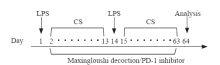
Figure 1.
Experimental design for COPD model and interventions.COPD:chronic obstructive pulmonary disease; LPS: lipopolysaccharide; CS: cigarette smoke.
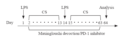

Figure 2.
H&E staining and IHC staining of lung tissue (left×20, right×40). IHC: immunohistochemistry. Group A: normal group; group B: COPD model group; group C: Maxingloushi decoction + COPD group; group D: PD-1 inhibitor + COPD group.
Table 1
IOD value of analysis of PD-1 and PD-L1 stained by IHC (mean±SD)
| Groups | n | PD-1 | PD-L1 |
|---|---|---|---|
| Group A | 9 | 386.26±85.41* | 37,193.11±7,382.04* |
| Group B | 9 | 27,748.31±7,532.47 | 4,159.88±389.25 |
| Group C | 9 | 3,484.48±922.72*# | 15,582.37±2,804.94*# |
| Group D | 9 | 4,527.86±1,122.84* | 7,807.50±2,660.21* |
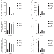
Figure 3.
Expression of PD-1 and PD-L1 in plasma and BLF. Compared with group B, *P<0.05; compared with group C, #P<0.05. Group A: normal group; group B: COPD model group; group C: Maxingloushi decoction + COPD group; group D: PD-1 inhibitor + COPD group; IOD: immuno-fluorescence optical density; IHC: immunohistochemistry; PD-1: programmed death-1; PD-L1: programmed death-ligand 1.

Table 2
Expression of PD-1 and PD-L1 in mouse plasma and bronchoalveolar lavage fluid (mean±SD, pg/mL)
| Groups | n | PD-1 (plasma) | PD-L1 (plasma) | PD-1 (bronchoalveolar lavage fluid) | PD-L1 (bronchoalveolar lavage fluid) |
|---|---|---|---|---|---|
| Group A | 6 | 132.65±4.43* | 14.76±1.13* | 88.24±5.90* | 6.92±0.69* |
| Group B | 10 | 416.56±8.80 | 2.52±0.62 | 295.20±18.12 | 1.43±0.25 |
| Group C | 10 | 388.50±11.87*# | 5.15±0.73*# | 252.33±6.24*# | 2.58±0.37*# |
| Group D | 10 | 401.83±10.95* | 3.52±0.90* | 275.21±14.15* | 1.66±0.42 |
| 1 | Liu ZY, Zhong NS. Internal Medicine (7th Edition). Beijing: People’s Medical Publishing House, 2008;62-3. |
| 2 |
Bi W, Sun Y, Ma LQ, Wu CJ. Predictive role of interleukin-6 and CAT score in mechanical ventilation in patients with chronic obstructive pulmonary disease at the acute exacerbation stage in the emergency department World J Emerg Med. 2020; 11(2):93-6.
doi: 10.5847/wjem.j.1920-8642.2020.02.005 |
| 3 | Mei HY, Niu HH, Li RX. Expression of SIRT1, NF-κB, and MMP-9 in peripheral blood of COPD patients. Medical Science Journal of Central South China. 2019; 47:281. |
| 4 | Hao Q, Yin YP, He M, Zhang LB, Zhu JY. Correlation between MCP-1, IL-17, IL-35 and lung function in patients with acute exacerbation of chronic obstructive pulmonary disease. Lab Immun Clin Med. 2020; 27(6):978-9. |
| 5 | Cha G, Wu XJ. The role of specific and non-specific immune cells in the pathogenesis of COPD Int J Respir. 2017; 37(16):1258-61. |
| 6 | Tong H, Wang YG. Progress in immunopathology of PD-1/PD-L1 axis and chronic obstructive pulmonary disease Int J Respir. 2020; 40(11):861-5. |
| 7 |
Zhang H, Sun D, Li D, Zheng Z, Xu J, Liang X, et al. Long non-coding RNA expression patterns in lung tissues of chronic cigarette smoke induced COPD mouse model. Sci Rep. 2018; 8(1):7609.
doi: 10.1038/s41598-018-25702-3 |
| 8 |
Li DF, Wang J, Sun DJ, Gong XF, Jiang H, Shu JZ, et al. Tanshinone IIA sulfonate protects against cigarette smoke-induced COPD and down-regulation of CFTR in mice. Sci Rep. 2018; 8(1):376.
doi: 10.1038/s41598-017-18745-5 |
| 9 |
Kuang J, Hou X, Zhang J, Chen Y, Su Z. Identification of insulin as a novel retinoic acid receptor-related orphan receptor α target gene. FEBS Lett. 2014; 588(6):1071-9.
doi: 10.1016/j.febslet.2014.02.029 |
| 10 |
Yang X, Huo B, Zhong XN, Su WY, Liu WT, Li YM, et al. Imbalance between subpopulations of regulatory T cells in patients with acute exacerbation of COPD. COPD. 2017; 14(6):618-25.
doi: 10.1080/15412555.2017.1385055 pmid: 29166179 |
| 11 | Li W, Wang S, Tian J, Wan Y. Role of regulatory T cells in the pathogenesis of COPD. Journal of Clinical Pulmonary Medicine. 2014; 19(1):128-9. |
| 12 | Wu WP. Collection of Wu Weiping’s Academic Thought and Clinical Experience. Beijing: China Traditional Chinese Medicine Publishing House, 2014; 70. |
| 13 | Yang T, Wang Q, Cheng Y. Effect of Maxingloushi decoction on acute AECOPD. Zhejiang Clinical Medical Journal. 2019; 21(5):621-2. |
| 14 | Li Z, Shi J, Li B. Effect of Maxingloushi decoction combined with budesonide atomization inhalation on AECOPD and its effect on Th1/Th2 imbalance. Chinese Journal of Coal Industry Medicine. 2019; 22(1):95-8. |
| 15 | Zhang J, Wu J. Inflammatory Factors and Chronic Disease Prevention and Control. Beijing: Science Press, 2019;2-3. |
| 16 | Zhou GY. Principles of Immunology. Beijing: Science Press, 2018;5-7. |
| 17 |
Lugade AA, Bogner PN, Thatcher TH, Sime PJ, Phipps RP, Thanavala Y. Cigarette smoke exposure exacerbates lung inflammation and compromises immunity to bacterial infection. J Immunol. 2014; 192(11):5226-35.
doi: 10.4049/jimmunol.1302584 pmid: 24752444 |
| 18 | Patsoukis N, Brown J, Petkova V, Liu F, Li L, Boussiotis VA. Selective effects of PD-1 on Akt and Ras pathways regulate molecular components of the cell cycle and inhibit T cell proliferation. Sci Signal. 2012; 5(230): ra46. |
| 19 | Wang X, Li W, Wang D, Song T, Yao Y. Effects of interleukin-37 on regulatory T cell immune function in sepsis mice. Chinese Journal of Emergency Medicine. 2020; 29(2):188-92. |
| [1] | Ren-qi Yao, Chao Ren, Di Ren, Jin-xiu Li, Ying Li, Xue-yan Liu, Lei Huang, Yong Liu, Mian Peng, Yong-wen Feng, Yong-ming Yao. Development of septic shock and prognostic assessment in critically ill patients with coronavirus disease outside Wuhan, China [J]. World Journal of Emergency Medicine, 2021, 12(4): 293-298. |
| [2] | Jun Yin, Yao Chen, Jun-ling Huang, Lei Yan, Zhong-shu Kuang, Ming-ming Xue, Si Sun, Hao Xiang, Yan-yan Hu, Zhi-min Dong, Chao-yang Tong, Chun-xue Bai, Zhen-ju Song. Prognosis-related classification and dynamic monitoring of immune status in patients with sepsis: A prospective observational study [J]. World Journal of Emergency Medicine, 2021, 12(3): 185-191. |
| [3] | Zhong-shu Kuang, Yi-lin Yang, Wei Wei, Jian-li Wang, Xiang-yu Long, Ke-yong Li, Chao-yang Tong, Zhan Sun, Zhen-ju Song. Clinical characteristics and prognosis of community-acquired pneumonia in autoimmune disease-induced immunocompromised host: A retrospective observational study [J]. World Journal of Emergency Medicine, 2020, 11(3): 145-151. |
| [4] | Wei Bi, Yan Sun, Lin-qin Ma, Cai-jun Wu. Predictive role of interleukin-6 and CAT score in mechanical ventilation in patients with chronic obstructive pulmonary disease at the acute exacerbation stage in the emergency department [J]. World Journal of Emergency Medicine, 2020, 11(2): 93-96. |
| [5] | Hong-wei Zhang, Li-you Wei, Gang Zhao, Ya-jing Yang, Shu-zheng Liu, Zhen-yu Zhang, Zhang Jing, Yan-ling Hu. Periplaneta americana extract used in patients with systemic inflammatory response syndrome [J]. World Journal of Emergency Medicine, 2016, 7(1): 50-54. |
| [6] | Şükrü Gürbüz, Mustafa Yıldız, Murat Kara, Kürşat Kargün, Mehtap Gürger, Metin Ateşçelik, Ömer Doğan Alataş. Paraoxonase-1 gene in patients with chronic obstructive pulmonary disease investigation Q192R and L55M polymorphisms [J]. World Journal of Emergency Medicine, 2015, 6(3): 201-206. |
| [7] | Jian-ping Gao, Ke-jing Ying. Thrombolysis during extended cardiopulmonary resuscitation for autoimmune-related pulmonary embolism [J]. World Journal of Emergency Medicine, 2015, 6(2): 153-156. |
| [8] | Chao Cao, Tao Ma, Yan-fen Chai, Song-tao Shou. The role of regulatory T cells in immune dysfunction during sepsis [J]. World Journal of Emergency Medicine, 2015, 6(1): 5-9. |
| [9] | Wei-ping Sun, Guang-xiong Yuan, Yan-juan Hu, Li-zhen Liao, Lin Fu. Effect of low-dose glucocorticoid on corticosteroid insufficient patients with acute exacerbation of chronic obstructive pulmonary disease [J]. World Journal of Emergency Medicine, 2015, 6(1): 34-39. |
| [10] | Xue-zhong Xing, Yong Gao, Hai-jun Wang, Quan-hui Yang, Chu-lin Huang, Shi-ning Qu, Hao Zhang, Hao Wang, Qing-ling Xiao, Ke-lin Sun. Risk factors and prognosis of critically ill cancer patients with postoperative acute respiratory insufficiency [J]. World Journal of Emergency Medicine, 2013, 4(1): 43-47. |
| [11] | Rong-rong Song, Yan-ping Qiu, Yong-ju Chen, Yong Ji. Application of fiberoptic bronchscopy in patients with acute exacerbations of chronic obstructive pulmonary disease during sequential weaning of invasive-noninvasive mechanical ventilation [J]. World Journal of Emergency Medicine, 2012, 3(1): 29-34. |
| [12] | Sha-luo Li, Yong-hua Xu, Xi Wang, Xue-feng Liu, Liang Zhao. Effects of enteral immunonutrition on immune function in patients with multiple trauma [J]. World Journal of Emergency Medicine, 2011, 2(3): 206-209. |
| [13] | Xing-hai Chen, Yong-jie Yin, Jing-xiao Zhang. Sepsis and immune response [J]. World Journal of Emergency Medicine, 2011, 2(2): 88-92. |
| [14] | Juan Xie, Xing-yi Yang, Jin-dong Shi, Xing-qi Deng, Wei Long. A new inflammation marker of chronic obstructive pulmonary disease- adiponectin [J]. World Journal of Emergency Medicine, 2010, 1(3): 190-195. |
| Viewed | ||||||
|
Full text |
|
|||||
|
Abstract |
|
|||||
