World Journal of Emergency Medicine ›› 2012, Vol. 3 ›› Issue (1): 60-64.doi: 10.5847/wjem.j.issn.1920-8642.2012.01.011
• Original Articles • Previous Articles Next Articles
Jing Shi1, Chun-lin Hu2, Yu-feng Gao1, Xiao-xing Liao2( ), Hope Xu3
Received:2011-07-27
Accepted:2011-12-29
Online:2012-03-15
Published:2012-03-15
Contact:
Xiao-xing Liao
E-mail:liaowens@163.com
Jing Shi, Chun-lin Hu, Yu-feng Gao, Xiao-xing Liao, Hope Xu. The relationship between platelet endothelial cell adhesion molecule-1 and paraquat-induced lung injury in rabbits[J]. World Journal of Emergency Medicine, 2012, 3(1): 60-64.
Add to citation manager EndNote|Ris|BibTeX
URL: http://wjem.com.cn//EN/10.5847/wjem.j.issn.1920-8642.2012.01.011
Table 1
The physiological variables of rabbits in each group
| Groups | BW (kg) | HR (Times/min) | RR (Times/min) | T (°C) |
|---|---|---|---|---|
| A | 2.41±0.26 | 236±36 | 32.3±2.34 | 38.5±2.34 |
| B | 2.45±0.23 | 245±56 | 29.8±4.56 | 38.2±1.78 |
| C | 2.38±0.46 | 239±27 | 30.4±5.26 | 39.1±1.05 |
| P value | 0.923 | 0.808 | 0.956 | 0.889 |
Table 2
The survival time of rabbits in each group
| Groups | Survival time (day) |
|---|---|
| A | 6.47±0.99 |
| B | 6.09±1.04 |
| C | 4.77±2.04*# |
| P value | 0.923 |
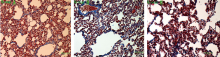
Figure 1.
The injury and fibrosis of the lungs in each group.
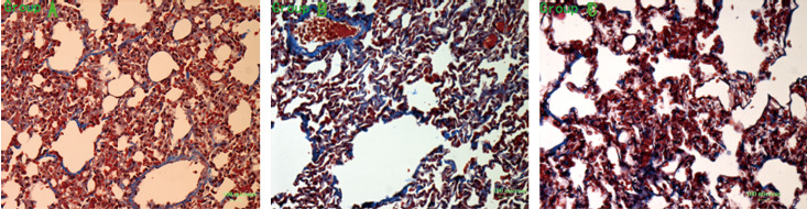
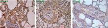
Figure 2.
The expression of PECAM-1 in the lungs in each group.

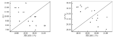
Figure 3.
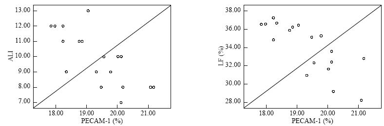
The correlation analysis of the PECAM-1 expression and ALI score and lung fibrosis.
| 1 |
Tsukamoto M, Tampo Y, Sawada M, Yonaha M. Paraquat-induced oxidative stress and dysfunction of the glutathione redox cycle in pulmonary microvascular endothelial cells. Toxicol Appl Pharmacol 2002; 178:82-92.
doi: 10.1006/taap.2001.9325 pmid: 11814328 |
| 2 |
Takizawa M, Komori K, Tampo Y, Yonaha M. Paraquat-induced oxidative stress and dysfunction of cellular redox systemsincluding antioxidative defense enzymes glutathione peroxidase and thioredoxinreductase. Toxicol In Vitro 2007; 21:355-363.
doi: 10.1016/j.tiv.2006.09.003 |
| 3 |
Tsukamoto M, Tampo Y, Sawada M, Yonaha M. Paraquat-induced membrane dysfunction in pulmonary microvascular endothelialcells. Pharmacol Toxicol 2000; 86:102-109.
doi: 10.1034/j.1600-0773.2000.d01-19.x pmid: 10752666 |
| 4 |
Takizawa M, Komori K, Tampo Y, Yonaha M. Paraquat-induced oxidative stress and dysfunction of cellular redox systemsincluding antioxidative defense enzymes glutathione peroxidase and thioredoxinreductase. Toxicol In Vitro 2007; 21:355-363.
doi: 10.1016/j.tiv.2006.09.003 |
| 5 |
Ahmad I, Kumar A, Shukla S, Prasad PH, Singh C. The involvement of nitric oxide in maneb- and paraquat-induced oxidative stressin rat polymorphonuclear leukocytes. Free Radic Res 2008; 42:849-862.
doi: 10.1080/10715760802513733 pmid: 18985485 |
| 6 |
Ahmad I, Shukla S, Kumar A. Maneb and paraquat-induced modulation of toxicant responsive genes in the ratliver: comparison with polymorphonuclear leukocytes. Chem Biol Interact 2010; 188:566-579.
doi: 10.1016/j.cbi.2010.09.023 pmid: 20888808 |
| 7 |
Kumar A, Ahmad I, Shukla S, Singh BK, Patel DK, Pandey HP. et al. Effect of zinc and paraquat co-exposure on neurodegeneration: Modulation ofoxidative stress and expression of metallothioneins, toxicant responsive andtransporter genes in rats. Free Radic Res 2010; 44:950-965.
pmid: 20553223 |
| 8 |
Suntres ZE. Role of antioxidants in paraquat toxicity. Toxicology 2002; 180:65-77.
doi: 10.1016/s0300-483x(02)00382-7 pmid: 12324200 |
| 9 |
Wang Y, Sheibani N. Expression pattern of alternatively spliced PECAM-1 isoforms in hematopoieticcells and platelets. J Cell Biochem 2002; 87:424-438.
doi: 10.1002/jcb.10321 pmid: 12397602 |
| 10 |
Lou MF. Redox regulation in the lens. Prog Retin Eye Res 2003; 22:657-682.
doi: 10.1016/s1350-9462(03)00050-8 pmid: 12892645 |
| 11 |
Wilhelm J, Smistik Z, Mahelkova G, Vytasek R. Redox regulation of proliferation of lens epithelial cells in culture. Cell Biochem Funct 2007; 25:317-321.
doi: 10.1002/cbf.1390 pmid: 17191273 |
| 12 |
Bonilla E, Medina-Leendertz S, Villalobos V, Molero L, Bohorquez A. Paraquat-induced oxidative stress in drosophila melanogaster: effects ofmelatonin, glutathione, serotonin, minocycline, lipoic acid and ascorbic acid. Neurochem Res 2006; 31:1425-1432.
doi: 10.1007/s11064-006-9194-8 |
| 13 |
Kumar A, Ahmad I, Shukla S, Singh BK, Patel DK, Pandey HP. et al. Endothelial cell activation and blood coagulation in critically ill patients withlung injury. Wien Klin Wochenschr. 2002; 114:853-858.
pmid: 12503477 |
| 14 |
Marszalek A, Daa T, Kashima K, Nakayama I, Yokoyama S. Ultrastructural and morphometric studies related to expression of the celladhesion molecule PECAM-1/CD31 in developing rat lung. J Histochem Cytochem 2000; 48:1283-1289.
doi: 10.1177/002215540004800911 pmid: 10950884 |
| 15 |
Carrithers M, Tandon S, Canosa S, Michaud M, Graesser D, Madri JA. Enhanced susceptibility to endotoxic shock and impaired STAT3 signaling inCD31-deficient mice. Am J Pathol 2005; 166:185-196.
doi: 10.1016/S0002-9440(10)62243-2 pmid: 15632011 |
| 16 |
Brown S, Heinisch I, Ross E, Shaw K, Buckley CD, Savill J. Apoptosis disables CD31-mediated cell detachment from phagocytes promotingbinding and engulfment. Nature 2002; 418:200-203.
doi: 10.1038/nature00811 pmid: 12110892 |
| 17 |
Wang Y, Sheibani N. Expression pattern of alternatively spliced PECAM-1 isoforms in hematopoieticcells and platelets. J Cell Biochem 2002; 87:424-438.
doi: 10.1002/jcb.10321 pmid: 12397602 |
| 18 |
Stalmans I. Role of the vascular endothelial growth factor isoforms in retinal angiogenesisand DiGeorge syndrome. Verh K Acad Geneeskd Belg 2005; 67:229-276.
pmid: 16334858 |
| [1] | Jian-hua Yi, Zhao-cai Zhang, Mei-bian Zhang, Xin He, Hao-ran Lin, Hai-wen Huang, Hai-bin Dai, Yu-wen Huang. Role of epithelial-to-mesenchymal transition in the pulmonary fibrosis induced by paraquat in rats [J]. World Journal of Emergency Medicine, 2021, 12(3): 214-220. |
| [2] | Yun-fei Jiang, Jian Kang, Pei-pei Huang, Jia-xi Yao, Zhong-he Wang, Lei Jiang, Jun Wang, Li Qiao, Bao-li Zhu, Hao Sun, Jin-song Zhang. Evaluation of gastric lavage efficiency and utility using a rapid quantitative method in a swine paraquat poisoning model [J]. World Journal of Emergency Medicine, 2020, 11(3): 174-181. |
| [3] | Jia-jun Xu, Jian-tao Zhen, Li Tang, Qing-ming Lin. Intravenous injection of Xuebijing attenuates acute kidney injury in rats with paraquat intoxication [J]. World Journal of Emergency Medicine, 2017, 8(1): 61-64. |
| [4] | Ming Wei, Yan-jie Gong, Ling Tu, Jia Li, Ying-hong Liang, Yi-hua Zhang. Expression of phosphatidylinositol-3 kinase and effects of inhibitor Wortmannin on expression of tumor necrosis factor-α in severe acute pancreatitis associated with acute lung injury [J]. World Journal of Emergency Medicine, 2015, 6(4): 299-304. |
| [5] | Zhi-wei Liu, Hai-ying Wang, Lan Guan, Bin Zhao. Regulatory effects of hydrogen sulfide on alveolar epithelial cell endoplasmic reticulum stress in rats with acute lung injury [J]. World Journal of Emergency Medicine, 2015, 6(1): 67-73. |
| [6] | Shou-peng Li, Ji-yuan Han, Peng Sun, Guo-yan Wu, Xiang-yan Bai. Effect of SP-A/B in lipoic acid on acute paraquat poisoning [J]. World Journal of Emergency Medicine, 2014, 5(1): 57-62. |
| [7] | Xiao-xiao Meng, Rui-lan Wang, Shan Gao, Hui Xie, Jiu-ting Tan, Yong-bin Qian. Effect of ulinastatin on paraquat-induced-oxidative stress in human type II alveolar epithelial cells [J]. World Journal of Emergency Medicine, 2013, 4(2): 133-137. |
| [8] | Yin-song Jiang, Yu-ying Ma, Zhan-qing Wang, Guang-jun Li. Therapeutic effects of smecta or smectite powder on rats with paraquat toxication [J]. World Journal of Emergency Medicine, 2013, 4(2): 144-150. |
| [9] | Zhi-jian Zhang, Li-bo Peng, Ya-juan Luo, Cong-yang Zhou. Prospective experimental studies on the renal protective effect of ulinastatin after paraquat poisoning [J]. World Journal of Emergency Medicine, 2012, 3(4): 299-304. |
| [10] | Zhi-qiang Cheng, Ji-yuan Han, Peng Sun, Yu-ying Weng, Jiao Chen, Guo-yan Wu, Hong-xia Ma. Edaravone attenuates paraquat-induced lung injury by inhibiting oxidative stress in human type II alveolar epithelial cells [J]. World Journal of Emergency Medicine, 2012, 3(1): 55-59. |
| [11] | Chang-bin Li, Xin-hua Li, Zhen Wang, Cheng-hua Jiang, Ai Peng. Serum paraquat concentration detected by spectrophotometry in patients with paraquat poisoning [J]. World Journal of Emergency Medicine, 2011, 2(3): 179-184. |
| [12] | Hui-li Zhang, Yuan-fei Liu, Xu-rui Luo, Wei-hua Tan, Liang Huang. Saturated hydrogen saline protects rats from acute lung injury induced by paraquat [J]. World Journal of Emergency Medicine, 2011, 2(2): 149-153. |
| [13] | Jian-qiang Wang, Chun Pan, Lin Liu, Liang Jin, Yi Yang, Hai-bo Qiu. Effect of post recruitment maneuver ventilation by different tidal volume on lung vascular endothelial diastole function in rats with acute lung injury [J]. World Journal of Emergency Medicine, 2011, 2(2): 141-148. |
| [14] | Xiao-li Xu, Wei Wang, Zu-jun Song, Hong Ding, Xiao-hong Duan, Huan-cheng Meng, Jian Chong. Imaging in detecting sites of pulmonary fibrosis induced by paraquat [J]. World Journal of Emergency Medicine, 2011, 2(1): 45-49. |
| [15] | Zhi-jian Zhang, Cong-yang Zhou, Ya-juan Luo, Hua-wei Xiong. Expression of heat shock protein 70 in lung tissues of acute paraquat poisoned rats and intervention of ulinastatin [J]. World Journal of Emergency Medicine, 2010, 1(3): 229-233. |
| Viewed | ||||||
|
Full text |
|
|||||
|
Abstract |
|
|||||
