Anatomical variation in the internal jugular vein: potential risk factors for central venous catheterization - a case report
|
Anatomical variation in the internal jugular vein: potential risk factors for central venous catheterization - a case report |
| Yidan Shan, Weijia Huang |
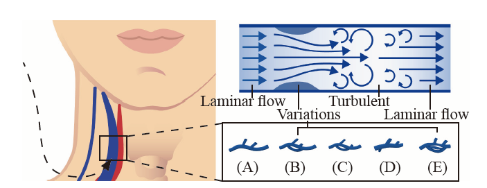
| Figure 1. Anatomical variation of the IJV and altered blood flow. A: normal IJV, which leads to laminar flow; B: duplication; C: fenestration; D: posterior tributary; E: trifurcation. B, C, D, and E may raise a turbulent blood flow due to the stenosis of the vessel theoretically. IJV: internal jugular vein. |
|
|