A case report of intramyocardial dissecting hematoma: a challenging diagnosis
|
A case report of intramyocardial dissecting hematoma: a challenging diagnosis |
| Yaoyao Zhu, Tong Wang, Longyuan Jiang, Lian Liang |
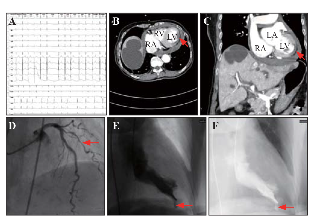
| Figure 1. Acute myocardial infarction complicated with intramyocardial dissecting hematoma. A: electrocardiogram showed atrial fibrillation with slight ST-segment elevation from V4 to V9; contrast-enhanced computed tomography (CT) scan of transverse section (B) and coronal section (C) revealed a low-enhanced segment in the anterior-lateral wall of the left ventricle (red arrows); D: coronary angiography found 70%-80% stenosis in the middle diagonal branch of the left coronary artery (red arrows); E and F: left ventricular angiography delineated a left ventricular subepicardial aneurysm characterized by an abrupt interruption of the myocardium (red arrow). LV: left ventricle; RV: right ventricle; RA: right atrium; LA: left atrium. |
|
|