Pregnancy-related spontaneous coronary artery dissection after intravenous ritodrine infusion: a case report
|
Pregnancy-related spontaneous coronary artery dissection after intravenous ritodrine infusion: a case report |
| Ya-qing An, Yan-ling Dong, Yi-jiao Men, Liang Liu, Yu Gong, Jian-ling Su, Heng-bo Gao, Ying-ping Tian |
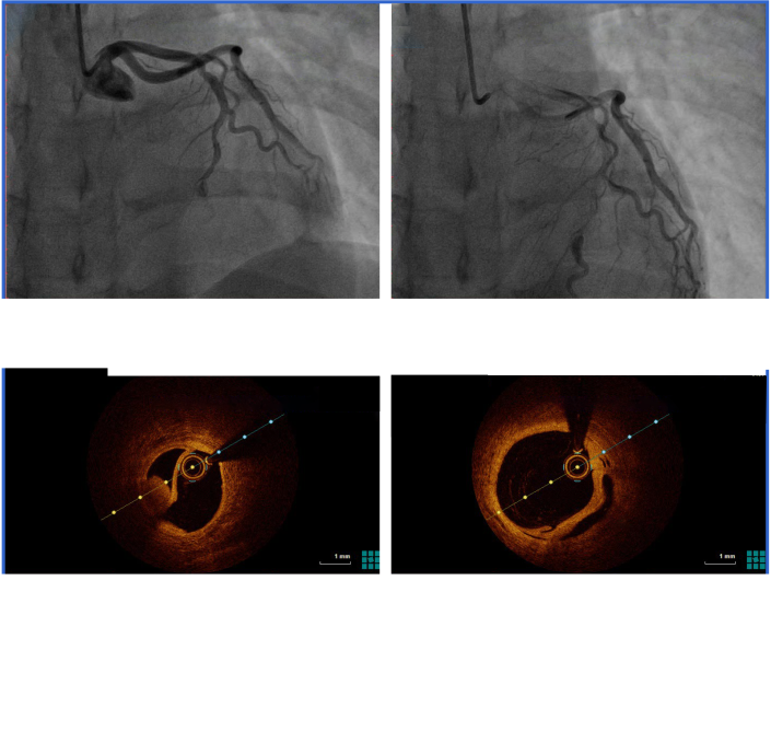
| Figure 2. Images of CAG and OCT. A, B: CAG showed an intimal tear (A, arrow) with persistent distal filling defects and a linear dissection (B, arrow) in the distal segment of LAD with false lumen filled with contrast agent; C, D: OCT indicated distal intimal detachment of LAD with intramural hematoma and thrombus (arrows). CAG: coronary angiography; OCT: optical coherence tomography; LAD: left anterior descending. |
|
|