A young male with abdominal distention and fever after cardiopulmonary resuscitation treatment
|
A young male with abdominal distention and fever after cardiopulmonary resuscitation treatment |
| Guo-feng Chen, Kai-bo Chen, Jian Chen |
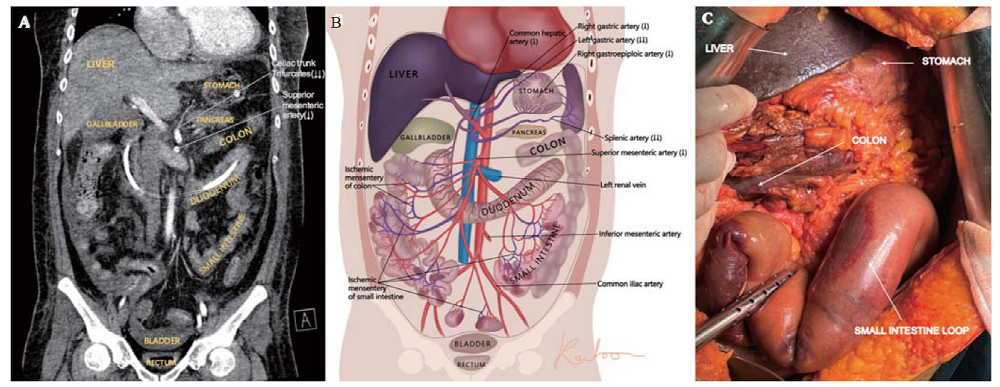
| Figure 2. Images of a computed tomography (CT) scan and intraoperative photograph. A: the contrast CT scan showed that there was no enhancement of the splenic artery and insufficient enhancement of the left gastric artery, the common hepatic artery and the superior mesenteric artery (shown in coronal view); B: the illustrative picture simulated different degrees of ischemia (↓: decreased blood flow; ↓↓: significantly decreased blood flow); C: intraoperative photograph demonstrated ischemia and necrosis of the entire perforated stomach, liver, and most segments of the small intestine and colon. |
|
|