A case of massive hemoptysis after radiofrequency catheter ablation for atrial fibrillation
|
A case of massive hemoptysis after radiofrequency catheter ablation for atrial fibrillation |
| Zhen Ren, Shu Li, Qing-bian Ma |
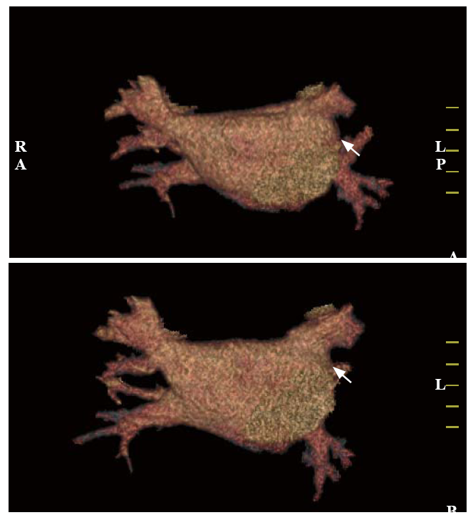
| Figure 2. Contrast-enhanced multi-slice CT venography with three-dimensional reconstruction. Both figures (A and B) revealed detected left superior PVS (shown by arrows). CT: computed tomography; PVS: pulmonary venous stenosis. |
|
|