An intramural left ventricular fistula caused by left ventriculography
|
An intramural left ventricular fistula caused by left ventriculography |
| Qian Nie, Haseeb Sattar, Ai-ling Huang, Hong-cai Zhang, Jue Zhao, Wen Xie |
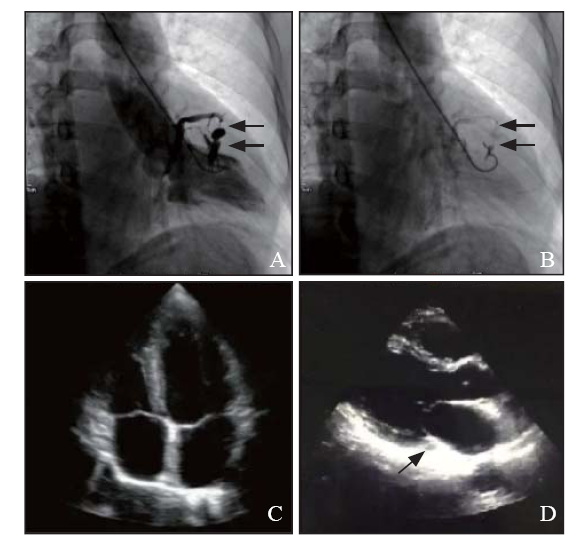
| Figure 1. Cardiac catheterization of left ventriculography and emergency echocardiography. A: intramural hematoma formed on the left ventricular wall caused by left ventriculography and several channels connected with coronary sinus (CS) (black arrows); B: re-ventriculography with lower pressure on the same position showed the potential fistula from left ventricle to CS (black arrows); C: 4-chamber echocardiography indicated no complication; D: no enlarged CS (black arrows) was found on the long axis of echocardiography. |
|
|