Point-of-care ultrasound identification of pneumatosis intestinalis associated with Henoch-Schönlein purpura gastrointestinal involvement: A case report
|
Point-of-care ultrasound identification of pneumatosis intestinalis associated with Henoch-Schönlein purpura gastrointestinal involvement: A case report |
| Sek Wan Tan, Vigil James, Aswin Warier, Gene Yong-kwang Ong |
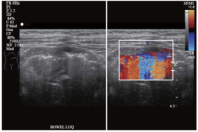
| Figure 2. Left upper quadrant ultrasound scan showing loops of thickened bowel with pneumatosis intestinalis (sub-serosal echogenic foci) and color flow Doppler study demonstrating significantly reduced blood flow in the bowel wall. Color flow Doppler artefacts can be observed arising from the intramural air. |
|
|