Point-of-care ultrasound identification of pneumatosis intestinalis associated with Henoch-Schönlein purpura gastrointestinal involvement: A case report
|
Point-of-care ultrasound identification of pneumatosis intestinalis associated with Henoch-Schönlein purpura gastrointestinal involvement: A case report |
| Sek Wan Tan, Vigil James, Aswin Warier, Gene Yong-kwang Ong |
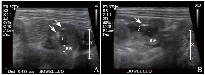
| Figure 1. Left upper quadrant longitudinal (A) and transverse (B) abdominal ultrasound scans showing a loop of thickened bowel wall. Presence of intramural air was identified by sub-serosal echogenic foci (arrows), with posterior echogenic shadows (arrowheads); BW: bowel wall; L: lumen (bowel). |
|
|