Effects of extracellular vesicles from mesenchymal stem cells on oxygen-glucose deprivation/reperfusion-induced neuronal injury
|
Effects of extracellular vesicles from mesenchymal stem cells on oxygen-glucose deprivation/reperfusion-induced neuronal injury |
| Shuang-shuang Gu, Xiu-wen Kang, Jun Wang, Xiao-fang Guo, Hao Sun, Lei Jiang, Jin-song Zhang |
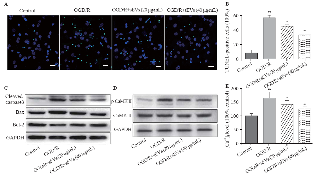
| Figure 3. Effects of BMSC-sEVs on OGD/R-induced neuronal apoptosis and Ca2+/CaMK II signaling pathway. Representative TUNEL images (A) and quantification of TUNEL-positive cells (B) (bar=20 μm); C: the protein levels of cleaved caspase-3, Bax and Bcl-2 determined by Western blot assay; D: the protein levels of CaMK II and p-CaMK II were determined by Western blot assay; E: the relative fluorescent intensity of Fluo-4 used to indicate the [Ca2+]i quantity; BMSCs: bone marrow mesenchymal stem cells; sEVs: small extracellular vesicles; OGD/R: oxygen-glucose deprivation and reperfusion; compared with control group, ##P<0.01; compared with OGD/R group, * P<0.05, **P<0.01. |
|
|