Effects of extracellular vesicles from mesenchymal stem cells on oxygen-glucose deprivation/reperfusion-induced neuronal injury
|
Effects of extracellular vesicles from mesenchymal stem cells on oxygen-glucose deprivation/reperfusion-induced neuronal injury |
| Shuang-shuang Gu, Xiu-wen Kang, Jun Wang, Xiao-fang Guo, Hao Sun, Lei Jiang, Jin-song Zhang |
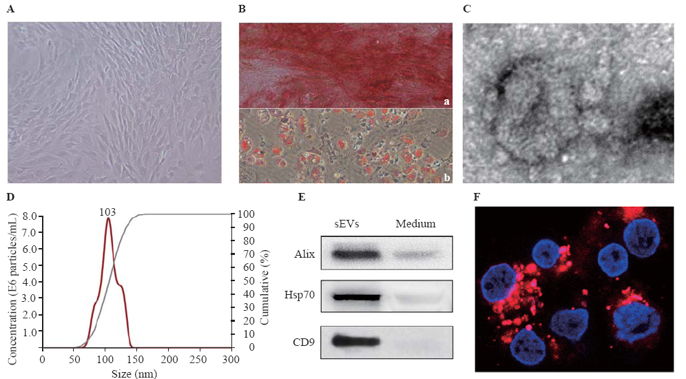
| Figure 1. Identification of BMSCs and BMSC-sEVs. A: morphology of BMSCs under phase-contrast microscope; B: multi-differentiation potential of BMSCs; a: Alizarin Red staining of osteogenic mineralization; b: Oil Red O staining of small lipid droplets (bar=100 μm); C: transmission electron micrograph of BMSC-sEVs (bar=50 nm); D: nanoparticle tracking analysis of sEVs; E: the expression of Alix, Hsp70, CD9 in conditioned medium and sEVs by Western blot assay; F: the uptake of PKH26-labeled sEVs (red) by neurons (4',6-diamidino-2-phenylindole [DAPI] blue) at 24 hours (bar=20 μm); BMSCs: bone marrow mesenchymal stem cells; sEVs: small extracellular vesicles. |
|
|