World Journal of Emergency Medicine ›› 2022, Vol. 13 ›› Issue (5): 349-354.doi: 10.5847/wjem.j.1920-8642.2022.080
• Original Articles • Previous Articles Next Articles
Gan-nan Wang1, Zhong-man Zhang1, Wen Chen2, Xiao-quan Xu2, Jin-song Zhang1( )
)
Received:2021-11-29
Accepted:2022-05-18
Online:2022-08-23
Published:2022-09-01
Contact:
Jin-song Zhang
E-mail:zhangjso@njmu.edu.cn
Gan-nan Wang, Zhong-man Zhang, Wen Chen, Xiao-quan Xu, Jin-song Zhang. Timing of brain computed tomography for predicting neurological prognosis in comatose cardiac arrest survivors: a retrospective observational study[J]. World Journal of Emergency Medicine, 2022, 13(5): 349-354.
Add to citation manager EndNote|Ris|BibTeX
URL: http://wjem.com.cn/EN/10.5847/wjem.j.1920-8642.2022.080
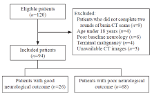
Figure 1.
Flow chart of patient enrollment.
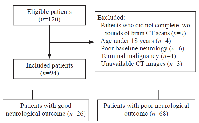
Table 1.
Clinical characteristic of enrolled patients
| Parameters | Good-outcome group (n=26) | Poor-outcome group (n=68) | P-value |
|---|---|---|---|
| Age, years | 52.62±16.30 | 49.72±19.93 | 0.511 |
| Male | 17 (65.4) | 47 (69.1) | 0.728 |
| Comorbidities | |||
| Hypertension | 12 (46.2) | 27 (39.7) | 0.570 |
| Diabetes | 6 (23.1) | 9 (13.2) | 0.395 |
| Coronary artery disease | 2 (7.7) | 9 (13.2) | 0.697 |
| Stroke | 3 (11.5) | 7 (10.3) | 0.861 |
| Out-of-hospital CA | 13 (50.0) | 38 (55.9) | 0.609 |
| Bystander CPR | 23 (88.5) | 44 (64.7) | 0.023 |
| Cardiac etiology | 15 (57.7) | 31 (45.6) | 0.294 |
| Initial shockable rhythm | 12 (46.2) | 20 (29.4) | 0.125 |
| Duration of resuscitation, min | 33.0 (19.0-45.5) | 30.0 (15.5-57.5) | 0.679 |
| GCS score after ROSC | 7.42±3.71 | 3.35±0.93 | <0.001 |
| Length of stay in hospital, d | 17.5 (14.0-28.5) | 9.0 (3.5-25.0) | 0.010 |
| CPC at hospital discharge | |||
| CPC 1 | 5 | - | |
| CPC 2 | 21 | - | |
| CPC 3 | - | 4 | |
| CPC 4 | - | 30 | |
| CPC 5 | - | 34 |
Table 2.
Comparison of CT parameters between different outcome groups by early (within 24 h) and late (24 h to 7 d) CTs
| Parameters | Early CT (< 24 h) | Late CT (24 h to 7 d) | ||||
|---|---|---|---|---|---|---|
| Good-outcome group (n=26) | Poor-outcome group (n=68) | P-value | Good-outcome group (n=26) | Poor-outcome group (n=68) | P-value | |
| Density of ROI, HU | ||||||
| PU | 34.1 (33.0-36.0) | 32.1 (28.2-35.9) | 0.078 | 33.5 (28.3-35.8) | 29.0 (26.0-32.0) | 0.028 |
| PLIC | 27.0 (24.4-30.0) | 28.0 (22.6-29.7) | 0.880 | 25.5 (21.3-26.9) | 25.0 (22.5-28.0) | 0.388 |
| GWR | ||||||
| Basal ganglia | 1.22 (1.16-1.29) | 1.17 (1.10-1.23) | 0.032 | 1.28 (1.23-1.35) | 1.12 (1.08-1.18) | <0.001 |
| Simplified | 1.24 (1.21-1.35) | 1.18 (1.12-1.27) | 0.052 | 1.33 (1.25-1.40) | 1.14 (1.08-1.22) | <0.001 |
| pCSFV | 0.040 (0.034-0.077) | 0.048 (0.030-0.070) | 0.824 | 0.074 (0.047-0.107) | 0.038 (0.019-0.075) | 0.010 |
Figure 2.
Comparison between different outcome groups in putamen density ratio (A), GWR ratio (B), and pCSFV ratio (C). GWR: grey-white matter ratio; pCSFV: proportion of cerebrospinal fluid volume.

Table 3.
ROC-analysis on the prediction of poor neurological outcome
| Prognostic factors | Cut-off value | Sensitivity (%) | Specificity (%) | AUC (95% CI) | P-value |
|---|---|---|---|---|---|
| GWR-basal ganglia (early CT) | 1.18 | 61.0 | 68.8 | 0.684 (0.542-0.826) | 0.032 |
| 1.12 | 31.7 | 100.0 | |||
| GWR-basal ganglia (late CT) | 1.18 | 87.8 | 93.8 | 0.942 (0.884-0.998) | <0.001 |
| 1.16 | 75.6 | 100.0 | |||
| GWR ratio | 1.01 | 78.0 | 75.0 | 0.761 (0.614-0.907) | 0.002 |
| pCSFV (late CT) | 0.03 | 41.5 | 100.0 | 0.721 (0.591-0.851) | 0.010 |
| pCSFV ratio | 0.98 | 65.9 | 93.8 | 0.788 (0.671-0.906) | 0.001 |
| 1 |
Gu SS, Kang XW, Wang J, Guo XF, Sun H, Jiang L, et al. Effects of extracellular vesicles from mesenchymal stem cells on oxygen-glucose deprivation/reperfusion-induced neuronal injury. World J Emerg Med. 2021; 12(1): 61-7.
doi: 10.5847/wjem.j.1920-8642.2021.01.010 |
| 2 | Virani SS, Alonso A, Benjamin EJ, Bittencourt MS, Callaway CW, Carson AP, et al. Heart disease and stroke statistics-2020update:a report from the American Heart Association. Circulation. 2020; 141(9): e139-596. |
| 3 |
Shao WJ, Shu TT, Xu S, Liang LC, Grange JML, Zhou YR, et al. Left-sided vagus nerve stimulation improves cardiopulmonary resuscitation outcomes in rats as effectively as right-sided vagus nerve stimulation. World J Emerg Med. 2021; 12(4): 309-16.
doi: 10.5847/wjem.j.1920-8642.2021.04.010 |
| 4 |
Sandroni C, D’Arrigo S, Nolan JP. Prognostication after cardiac arrest. Crit Care. 2018; 22(1): 150.
doi: 10.1186/s13054-018-2060-7 |
| 5 |
Greer DM, Wu O. Neuroimaging in cardiac arrest prognostication. Semin Neurol. 2017; 37(1): 66-74.
doi: 10.1055/s-0036-1594253 |
| 6 |
Keijzer HM, Hoedemaekers CWE, Meijer FJA, Tonino BAR, Klijn CJM, Hofmeijer J. Brain imaging in comatose survivors of cardiac arrest: Pathophysiological correlates and prognostic properties. Resuscitation. 2018; 133: 124-36.
doi: S0300-9572(18)30889-X pmid: 30244045 |
| 7 |
Lopez Soto C, Dragoi L, Heyn CC, Kramer A, Pinto R, Adhikari NKJ, et al. Imaging for neuroprognostication after cardiac arrest: systematic review and meta-analysis. Neurocrit Care. 2020; 32(1): 206-16.
doi: 10.1007/s12028-019-00842-0 |
| 8 |
Taccone FS, Baar I, de Deyne C, Druwe P, Legros B, Meyfroidt G, et al. Neuroprognostication after adult cardiac arrest treated with targeted temperature management: task force for Belgian recommendations. Acta Neurol Belg. 2017; 117(1): 3-15.
doi: 10.1007/s13760-017-0755-1 pmid: 28168412 |
| 9 |
You YH, Park JS, Yoo IS, Min JH, Jeong WJ, Cho YC, et al. Usefulness of a quantitative analysis of the cerebrospinal fluid volume proportion in brain computed tomography for predicting neurological prognosis in cardiac arrest survivors who undergo target temperature management. J Crit Care. 2019; 51: 170-4.
doi: 10.1016/j.jcrc.2019.02.024 |
| 10 |
Streitberger KJ, Endisch C, Ploner CJ, Stevens R, Scheel M, Kenda M, et al. Timing of brain computed tomography and accuracy of outcome prediction after cardiac arrest. Resuscitation. 2019; 145: 8-14.
doi: S0300-9572(19)30636-7 pmid: 31585185 |
| 11 |
Langkjær S, Hassager C, Kjaergaard J, Salam I, Thomsen JH, Lippert FK, et al. Prognostic value of reduced discrimination and oedema on cerebral computed tomography in a daily clinical cohort of out-of-hospital cardiac arrest patients. Resuscitation. 2015; 92: 141-7.
doi: 10.1016/j.resuscitation.2015.03.023 pmid: 25882783 |
| 12 |
Langhelle A, Nolan J, Herlitz J, Castren M, Wenzel V, Soreide E, et al. Recommended guidelines for reviewing, reporting, and conducting research on post-resuscitation care: the Utstein style. Resuscitation. 2005; 66(3): 271-83.
pmid: 16129543 |
| 13 |
Wang GN, Chen XF, Lv JR, Sun NN, Xu XQ, Zhang JS. The prognostic value of gray-white matter ratio on brain computed tomography in adult comatose cardiac arrest survivors. J Chin Med Assoc. 2018; 81(7): 599-604.
doi: 10.1016/j.jcma.2018.03.003 |
| 14 |
Chen YS, Dhar R, Heitsch L, Ford A, Fernandez-Cadenas I, Carrera C, et al. Automated quantification of cerebral edema following hemispheric infarction: application of a machine-learning algorithm to evaluate CSF shifts on serial head CTs. Neuroimage Clin. 2016; 12: 673-80.
doi: 10.1016/j.nicl.2016.09.018 |
| 15 |
Ajam K, Gold LS, Beck SS, Damon S, Phelps R, Rea TD. Reliability of the Cerebral Performance Category to classify neurological status among survivors of ventricular fibrillation arrest: a cohort study. Scand J Trauma Resusc Emerg Med. 2011; 19: 38.
doi: 10.1186/1757-7241-19-38 |
| 16 |
Sandroni C, Cariou A, Cavallaro F, Cronberg T, Friberg H, Hoedemaekers C, et al. Prognostication in comatose survivors of cardiac arrest: an advisory statement from the European Resuscitation Council and the European Society of Intensive Care Medicine. Resuscitation. 2014; 85(12): 1779-89.
pmid: 25438253 |
| 17 | Callaway CW, Donnino MW, Fink EL, Geocadin RG, Golan E, Kern KB, et al. Part 8: post-cardiac arrest care: 2015 American Heart Association guidelines update for cardiopulmonary resuscitation and emergency cardiovascular care. Circulation. 2015; 132(18 Suppl 2): S465-82. |
| 18 |
Hong JY, Lee DH, Oh JH, Lee SH, Choi YH, Kim SH, et al. Grey-white matter ratio measured using early unenhanced brain computed tomography shows no correlation with neurological outcomes in patients undergoing targeted temperature management after cardiac arrest. Resuscitation. 2019; 140: 161-9.
doi: 10.1016/j.resuscitation.2019.03.039 |
| 19 |
Moseby-Knappe M, Pellis T, Dragancea I, Friberg H, Nielsen N, Horn J, et al. Head computed tomography for prognostication of poor outcome in comatose patients after cardiac arrest and targeted temperature management. Resuscitation. 2017; 119: 89-94.
doi: S0300-9572(17)30270-8 pmid: 28687281 |
| 20 | Selip DB, Jantzie LL, Chang M, Jackson MC, Fitzgerald EC, Boll G, et al. Regional differences in susceptibility to hypoxic-ischemic injury in the preterm brain: exploring the spectrum from white matter loss to selective grey matter injury in a rat model. Neurol Res Int. 2012; 2012: 725184. |
| 21 |
Fujioka M, Okuchi K, Sakaki T, Hiramatsu K, Miyamoto S, Iwasaki S. Specific changes in human brain following reperfusion after cardiac arrest. Stroke. 1994; 25(10): 2091-5.
pmid: 8091457 |
| 22 |
Hayman EG, Patel AP, Kimberly WT, Sheth KN, Simard JM. Cerebral edema after cardiopulmonary resuscitation: a therapeutic target following cardiac arrest? Neurocrit Care. 2018; 28(3): 276-87.
doi: 10.1007/s12028-017-0474-8 pmid: 29080068 |
| 23 |
Minnerup J, Broocks G, Kalkoffen J, Langner S, Knauth M, Psychogios MN, et al. Computed tomography-based quantification of lesion water uptake identifies patients within 4.5 hours of stroke onset: a multicenter observational study. Ann Neurol. 2016; 80(6): 924-34.
doi: 10.1002/ana.24818 pmid: 28001316 |
| 24 |
Endisch C, Westhall E, Kenda M, Streitberger KJ, Kirkegaard H, Stenzel W, et al. Hypoxic-ischemic encephalopathy evaluated by brain autopsy and neuroprognostication after cardiac arrest. JAMA Neurol. 2020; 77(11): 1430-9.
doi: 10.1001/jamaneurol.2020.2340 |
| [1] | Fei Shao, Xian Shi, Shu-hua Huo, Qing-yu Liu, Ji-xue Shi, Jian Kang, Ping Gong, Sheng-tao Yan, Guo-xing Wang, Li-jie Qin, Fei Wang, Ke Feng, Feng-ying Chen, Yong-jie Yin, Tao Ma, Yan Li, Yang Wu, Hao Cui, Chang-xiao Yu, Song Yang, Wei Gan, Sai Wang, Liu-ye-zi Du, Ming-chen Zhao, Zi-ren Tang, Shen Zhao. Development and evaluation of a predictive nomogram for survival in heat stroke patients: a retrospective cohort study [J]. World Journal of Emergency Medicine, 2022, 13(5): 355-360. |
| [2] | Shi-jiao Yan, Mei Chen, Jing Wen, Wen-ning Fu, Xing-yue Song, Huan-jun Chen, Ri-xing Wang, Mei-ling Chen, Xiao-tong Han, Chuan-zhu Lyu. Global research trends in cardiac arrest research: a visual analysis of the literature based on CiteSpace [J]. World Journal of Emergency Medicine, 2022, 13(4): 290-296. |
| [3] | Hong-li Xiao, Lian-xing Zhao, Jun Yang, Nan Tong, Le An, Guo-xing Wang, Miao-rong Xie, Chun-sheng Li. Increasing angiotensin-converting enzyme (ACE) 2/ACE axes ratio alleviates early pulmonary vascular remodeling in a porcine model of acute pulmonary embolism with cardiac arrest [J]. World Journal of Emergency Medicine, 2022, 13(3): 208-214. |
| [4] | Chao-yu Lei, Heng-wei Qin, Xue-jie Dong, Jia-lin You, Lin Zhang. Layperson’s performance on an unconversant type of AED device: A prospective crossover simulation experimental study [J]. World Journal of Emergency Medicine, 2022, 13(2): 98-105. |
| [5] | Ji-yang Ling, Chun-sheng Li, Yun Zhang, Xiao-li Yuan, Bo Liu, Yong Liang, Qiang Zhang. Protective effect of extracorporeal membrane pulmonary oxygenation combined with cardiopulmonary resuscitation on post-resuscitation lung injury [J]. World Journal of Emergency Medicine, 2021, 12(4): 303-308. |
| [6] | Wei-jing Shao, Ting-ting Shu, Shuang Xu, Li-cai Liang, Jehane Michael Le Grange, Yu-ran Zhou, He Huang, Yu Cai, Qing Zhang, Peng Sun. Left-sided vagus nerve stimulation improves cardiopulmonary resuscitation outcomes in rats as effectively as right-sided vagus nerve stimulation [J]. World Journal of Emergency Medicine, 2021, 12(4): 309-316. |
| [7] | Yu-ming Wang, Yan-jun Zheng, Ying Chen, Yun-chuan Huang, Wei-wei Chen, Ran Ji, Li-li Xu, Zhi-tao Yang, Hui-qiu Sheng, Hong-ping Qu, En-qiang Mao, Er-zhen Chen. Effects of fluid balance on prognosis of acute respiratory distress syndrome patients secondary to sepsis [J]. World Journal of Emergency Medicine, 2020, 11(4): 216-222. |
| [8] | Xue-jie Dong, Lin Zhang, Yue-lin Yu, Shu-xiao Shi, Xiao-chen Yang, Xiao-qian Zhang, Shuang Tian, Helge Myklebust, Guo-hong Li, Zhi-jie Zheng. The general public’s ability to operate automated external defibrillator: A controlled simulation study [J]. World Journal of Emergency Medicine, 2020, 11(4): 238-245. |
| [9] | Wen-peng Yin, Jia-bao Li, Xiao-fang Zheng, Le An, Huan Shao, Chun-sheng Li. Effect of neutrophil CD64 for diagnosing sepsis in emergency department [J]. World Journal of Emergency Medicine, 2020, 11(2): 79-86. |
| [10] | Ye-cheng Liu, Yan-meng Qi, Hui Zhang, Joseph Walline, Hua-dong Zhu. A survey of ventilation strategies during cardiopulmonary resuscitation [J]. World Journal of Emergency Medicine, 2019, 10(4): 222-227. |
| [11] | Alexei Birkun, Yekaterina Kosova. Social attitude and willingness to attend cardiopulmonary resuscitation training and perform resuscitation in the Crimea [J]. World Journal of Emergency Medicine, 2018, 9(4): 237-248. |
| [12] | Alexei Birkun, Maksim Glotov, Herman Franklin Ndjamen, Esther Alaiye, Temidara Adeleke, Sergey Samarin. Pre-recorded instructional audio vs. dispatchers’ conversational assistance in telephone cardiopulmonary resuscitation: A randomized controlled simulation study [J]. World Journal of Emergency Medicine, 2018, 9(3): 165-171. |
| [13] | Shahin Shadnia, Nasim Zaman, Hossein Hassanian-Moghaddam, Hamed Shafaroodi, Mina Padandar, Mohammad Hasan Rezaeizadeh. Prognostic value of cortisol and thyroid function tests in poisoned patients admitted to toxicology ICU [J]. World Journal of Emergency Medicine, 2018, 9(1): 51-55. |
| [14] | Alexei Birkun, Maksim Glotov. Education in cardiopulmonary resuscitation in Russia: A systematic review of the available evidence [J]. World Journal of Emergency Medicine, 2017, 8(4): 245-252. |
| [15] | Chennappa Kalvatala Krishna, Hakim Irfan Showkat, Meenakshi Taktani, Vikram Khatri. Out of hospital cardiac arrest resuscitation outcome in North India — CARO study [J]. World Journal of Emergency Medicine, 2017, 8(3): 200-205. |
| Viewed | ||||||
|
Full text |
|
|||||
|
Abstract |
|
|||||
